
Most people assume skin imaging is just a high-resolution photo. What is spectral imaging skin analysis, though, goes far beyond that. Instead of capturing how your skin looks, spectral imaging captures what your skin is made of at a biochemical level, revealing pigmentation, vascular structures, and oxidative stress that no standard camera can detect. If you are researching advanced skin analysis techniques to make smarter skincare decisions or understand what dermatologists actually see, this guide breaks down exactly how the technology works, what its real-world applications are, and where its limits currently lie.
Table of Contents
- Key takeaways
- What is spectral imaging skin analysis?
- Lighting modes and skin analysis techniques
- Benefits and limitations of spectral imaging
- Comparing spectral imaging system types
- Practical applications in skincare and dermatology
- My honest take on where spectral imaging stands
- How Melaninglow uses spectral analysis for your skin
- FAQ
Key takeaways
| Point | Details |
|---|---|
| Beyond surface photography | Spectral imaging captures biochemical and structural skin data invisible to the naked eye, not just surface appearance. |
| Two main system types | Multispectral imaging suits portable, real-time devices; hyperspectral imaging provides deeper biochemical detail at higher complexity. |
| Multiple light modes matter | UV, cross-polarized, and standard light modes each reveal different skin features, from porphyrins to subsurface redness. |
| Adjunct tool, not a diagnosis | Spectral imaging currently supports dermatological assessment but cannot replace clinical diagnosis without further validation. |
| AI is expanding its reach | AI and physics-guided modeling are making spectral data analysis faster, more accurate, and increasingly accessible. |
What is spectral imaging skin analysis?
Standard digital cameras capture three color channels: red, green, and blue. Spectral imaging breaks light into dozens or even hundreds of narrow wavelength bands, creating what researchers call a spectral data cube. Every pixel in that cube holds a full spectrum of light information, not just a color value. Spectral imaging systems capture high-dimensional data cubes requiring calibration and correction, but the payoff is extraordinary: you can extract the biochemical fingerprints of skin chromophores like melanin, hemoglobin, and collagen directly from the image data.
How does spectral imaging work in practice? The process follows four core steps:
- Illumination: Controlled light sources illuminate the skin across specific wavelength ranges, from visible to near-infrared.
- Capture: A specialized sensor records how the skin reflects, absorbs, or transmits light at each wavelength band.
- Calibration: Raw spectral data is corrected against reference standards to remove sensor noise and lighting inconsistencies.
- Analysis: Software models decompose the spectral data into maps of chromophore concentrations, texture features, or lesion characteristics.
The key distinction within spectral imaging technology is the difference between multispectral and hyperspectral systems. Hyperspectral imaging captures 100 to 300+ narrow bands while multispectral uses fewer, broader bands. Hyperspectral systems produce richer biochemical detail but generate enormous data volumes and require more processing time. Multispectral systems trade some of that detail for speed and portability, making them better suited for clinical and consumer devices.
Pro Tip: When evaluating any spectral skin analysis device, ask specifically how many wavelength bands it captures and whether it uses multispectral or hyperspectral technology. That single answer tells you a lot about what the system can and cannot detect.
Lighting modes and skin analysis techniques
The wavelength bands a system uses are only part of the story. The type of illumination applied to the skin changes what you can see just as dramatically. Advanced skin analysis systems combine several lighting modes to build a complete picture of skin health.
- Standard white light imaging captures the skin surface as the eye sees it, revealing texture, pores, fine lines, and surface pigmentation. It is the baseline mode for any skin imaging session.
- Cross-polarized light filters out surface reflections to reveal what lies beneath the skin's outer layer. This mode is particularly effective for assessing redness, rosacea, and subsurface vascular structures that standard light obscures.
- UV fluorescence imaging uses ultraviolet light to excite fluorescent compounds in the skin. UV fluorescence in systems like VISIA reveals porphyrins produced by bacteria in pores, as well as early sun damage that has not yet surfaced visually.
- Near-infrared imaging penetrates deeper into the dermis, providing information about collagen density, hydration, and deeper vascular networks.
- RBX technology separates red and brown skin components to isolate vascular and pigmentation abnormalities like spider veins and hyperpigmentation with precision that standard color imaging cannot match.
Each mode answers a different clinical question. A single-mode device gives you one answer. A system that combines all five gives you a map of your skin's biology from the surface down to the dermis.
Benefits and limitations of spectral imaging
The benefits of skin analysis using spectral imaging are significant, particularly when compared to visual inspection or standard photography. Here is what the technology genuinely delivers.
What spectral imaging does well:
- It is entirely non-invasive. No biopsies, no dyes, no physical contact required beyond placing the skin near the sensor.
- Spectral data detects changes before visual signs appear, making it a powerful tool for early detection of pigmentation disorders and oxidative stress.
- Quantitative metrics allow you to track skin condition changes over time with numbers, not just subjective impressions.
- Multispectral imaging extracts chromophore concentration maps that support early melanoma screening as an adjunct tool.
Where the technology currently falls short:
- Acquisition variability affects result reliability significantly. Small changes in lighting angle, skin pressure, or calibration between sessions can confound results, especially in longitudinal tracking.
- Two devices using similar hardware can produce different biomarker maps if their chromophore models or illumination setups differ, which complicates cross-device comparisons.
- Spectral imaging remains an adjunct diagnostic tool, not a standalone diagnostic method. Clinical validation and standardization are still catching up to the technology's capabilities.
Pro Tip: If you are using spectral imaging for longitudinal skin monitoring, the consistency of your imaging conditions matters as much as the device itself. Same time of day, same skin prep, same distance from the sensor. Variability in those factors is the most common reason tracking results look inconsistent.
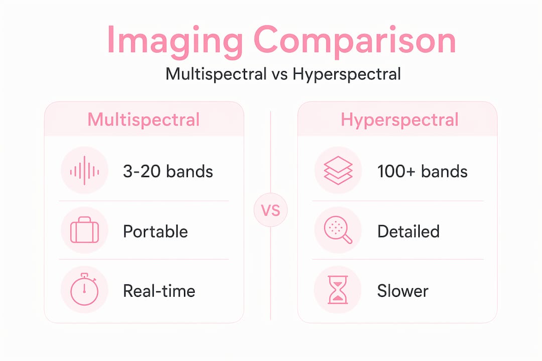
Comparing spectral imaging system types
Choosing between multispectral and hyperspectral imaging depends on what you need to measure and in what setting. The table below summarizes the practical trade-offs.
| Feature | Multispectral imaging | Hyperspectral imaging |
|---|---|---|
| Number of bands | 3 to 20 bands | 100 to 300+ bands |
| Spectral resolution | Lower | Higher |
| Data volume | Moderate | Very high |
| Acquisition speed | Fast, near real-time | Slower, scan-based |
| Device size | Compact, portable | Larger, lab-grade |
| Cost | Lower | Higher |
| Best use case | Clinical, aesthetic, consumer | Research, detailed biochemical analysis |
| Biochemical detail | Chromophore maps, key markers | Full spectral fingerprinting |
The trade-off between hyperspectral and multispectral systems determines whether a device can operate in real-time as a compact unit or provide detailed mechanistic biomarker quantification in a research setting. For most skincare and clinical aesthetic applications, multispectral snapshot devices hit the right balance. For dermatology research investigating the spectral signatures of rare lesions or novel chromophores, hyperspectral systems remain the gold standard.

Commercial devices like Canfield's VISIA fall into the multispectral category, using a defined set of lighting modes and wavelength channels to deliver clinically actionable skin analysis at scale. Research-grade hyperspectral systems, by contrast, are typically scan-based, meaning they build the spectral cube line by line, which takes longer and requires the subject to remain completely still.
Practical applications in skincare and dermatology
Understanding what is skin imaging at a spectral level opens up a range of applications that go well beyond a flattering before-and-after photo. Here is where spectral imaging technology is making a real difference.
- Early melanoma screening: Hyperspectral imaging tracks melanoma and sunspots across the 440 to 900 nm range, enabling longitudinal monitoring of lesion changes before they become visually obvious. As an adjunct to dermoscopy, it adds a biochemical layer to visual assessment.
- Pigmentation and oxidative stress monitoring: Spectral imaging is sensitive to biochemical processes like melanin distribution and oxidative stress, detecting subtle shifts that standard photography misses entirely.
- Personalized skincare recommendations: Objective chromophore maps and texture data give skincare professionals and AI systems a factual basis for product recommendations, moving beyond skin type questionnaires to actual measured skin biology.
- Treatment outcome tracking: Clinicians use repeated spectral imaging sessions to measure whether a treatment is producing quantifiable changes in vascular redness, pigmentation, or collagen density, not just whether the patient feels it is working.
- AI-enhanced analysis: AI and physics-guided modeling are emerging as vital tools for managing hyperspectral data complexity, improving accuracy, and making spectral analysis feasible on mobile and consumer platforms. This is the direction the field is moving fastest.
The intersection of spectral imaging with AI is particularly significant for melanin-rich skin tones, where standard skin analysis tools have historically underperformed. Spectral data is inherently more objective than visual scoring systems, which means it has the potential to deliver accurate analysis across the full range of human skin tones when the underlying models are trained correctly.
My honest take on where spectral imaging stands
I have spent a lot of time looking at how spectral imaging is marketed versus what the research actually supports, and the gap is real. The technology is genuinely extraordinary. Capturing a full biochemical map of your skin from a non-invasive image is not science fiction. It is happening in research labs and clinical devices right now.
What I find gets glossed over is the calibration problem. Interpretation of spectral maps depends heavily on system calibration and data processing quality. When a device or app claims to give you a spectral skin analysis, the meaningful question is not whether it uses spectral technology. The question is whether the underlying model has been validated, whether calibration is rigorous, and whether the chromophore assumptions match the skin tones being analyzed.
For melanin-rich skin specifically, this matters enormously. Most spectral skin models have been developed and validated predominantly on lighter skin tones. A system that has not been trained on diverse skin data is going to produce chromophore maps that are less reliable for darker complexions, regardless of how sophisticated the hardware is.
What genuinely excites me is the trajectory. Miniaturization is making spectral sensors small enough for handheld devices. AI is making the data processing fast enough for real-time use. And the field is slowly, finally, starting to take skin tone diversity seriously in its validation datasets. The next five years in spectral skin analysis are going to look very different from the last ten.
My advice: look for systems that are transparent about their validation data, their calibration protocols, and their limitations. The ones worth trusting are the ones that tell you what they cannot do.
— Orinami
How Melaninglow uses spectral analysis for your skin

Melaninglow was built specifically for the skin tones that generic analysis tools get wrong. The platform uses AI-driven facial analysis with multiple imaging modes, including UV illumination, to detect pigmentation patterns, texture variations, and skin concerns with the kind of specificity that actually reflects the biology of melanin-rich skin. Every analysis is processed locally on your device, meaning your images are never stored or shared with third parties.
If you have been researching spectral imaging and want to experience what objective, data-driven skin analysis looks like in practice, Melaninglow gives you a starting point built around your actual skin, not a generic template. Try the AI facial analysis and see what your skin is telling you beyond the surface.
FAQ
What is spectral imaging skin analysis?
Spectral imaging skin analysis captures light reflected from skin across multiple wavelength bands to reveal biochemical and structural skin properties, including melanin distribution, vascular patterns, and oxidative stress, that standard cameras cannot detect.
How does spectral imaging work on skin?
A spectral imaging system illuminates the skin with controlled light, captures how the skin reflects that light at each wavelength, and uses software models to extract chromophore concentration maps and other skin biomarkers from the resulting data cube.
Is spectral skin analysis effective for detecting skin conditions?
Spectral imaging is effective as an adjunct tool for detecting pigmentation disorders, early lesion changes, and vascular abnormalities, but it currently requires clinical validation before it can be used as a standalone diagnostic method.
What is the difference between multispectral and hyperspectral skin imaging?
Multispectral imaging uses 3 to 20 wavelength bands and suits portable, real-time clinical devices, while hyperspectral imaging captures 100 to 300+ bands for deeper biochemical detail but requires more complex, lab-grade equipment.
Can spectral imaging work for darker skin tones?
Spectral imaging technology can work across all skin tones, but the accuracy of results depends on whether the underlying chromophore models were validated on diverse skin tones. Systems trained predominantly on lighter skin may produce less reliable biomarker maps for melanin-rich complexions.